

Earlier this month, significant advancements in space exploration were marked by the first-ever X-ray image taken in space, facilitated by Canadian technology. This milestone was achieved aboard the SpaceX Dragon capsule, which recently completed a three-and-a-half-day mission that included orbiting the north and south poles. The mission was chartered by bitcoin investor Chun Wang and launched from NASA's Kennedy Space Center on March 31, with three other tourists accompanying him.
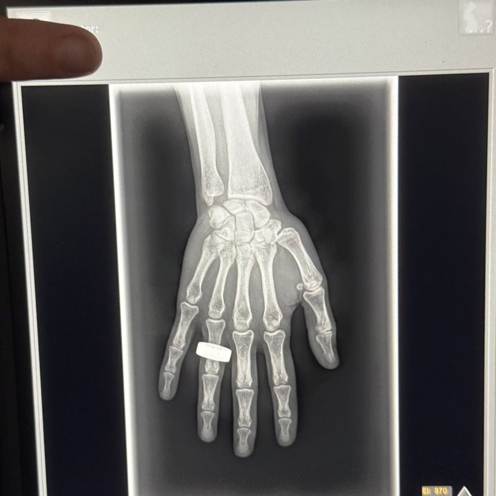
The X-ray image, featuring a hand with a ring, has been touted as a landmark achievement for Canadians in the realm of space exploration. Amol Karnick, CEO of KA Imaging, a company based in Waterloo, Ontario, expressed pride in this accomplishment, noting that the X-ray was produced using their innovative multi-image X-ray detector technology. KA Imaging's technology is recognized as the world's first of its kind, making this event particularly significant.
Karnick explained that this groundbreaking X-ray was part of the SpaceXray project, which was among 22 scientific studies selected for the mission. The ability to perform X-rays in space carries immense importance, as monitoring astronauts' bone mineral density is crucial due to the adverse effects of microgravity. The X-ray receiver developed by KA Imaging has shown the potential to mitigate those risks effectively.
Furthermore, Karnick highlighted that the X-ray receiver could serve dual purposes in space—diagnosing equipment issues or assessing the quality of repairs made on the spacecraft, which is vital for the safety and efficiency of space missions. The compact design of the KA Imaging X-ray receiver, comparable in size to a large laptop, also has advanced capabilities, allowing it to distinguish between various tissue types, such as bones and soft tissues like the lungs or heart. This functionality could prove invaluable for diagnosing conditions such as pneumonia or cancer.
Before the successful capture of the X-ray image, there were concerns about the feasibility of X-ray imaging in the space environment. Specifically, there were worries that the natural background radiation present in space might compromise the quality of the images. However, after reviewing the initial images received, Karnick confirmed that they exhibited no such issues, thereby affirming the viability of X-ray technology in space.
Notably, the crew aboard the spacecraft were not trained X-ray technicians, which necessitated that Karnick and his team train them on how to operate the equipment prior to the mission. In addition to the X-ray receiver, the setup included a portable X-ray generator produced by Chicago-based MinXray, further enhancing the mission's scientific capabilities.
Sheyna Gifford, the principal investigator of the SpaceXray study, expressed gratitude toward the crew, emphasizing the potential of this technology to improve safety and quality of life in space. Gifford also noted the aspiration for the same technological advancements to benefit life on Earth, particularly in remote or underserved areas, where access to advanced imaging may be limited.
This pioneering achievement sets the stage for further exploration into the practical applications of medical imaging technologies both in space and on our home planet. The collaboration between scientists and entrepreneurs from Canada underscores the important role that innovation plays in the future of space exploration and health care advancements.